The comprehensive journal Journal of Advanced Research published the latest article titled "Half-life determination of inorganic-organic hybrid nanomaterials in mice using laser-induced breakdown spectroscopy" by associate professor Guo Lianbo of Wuhan National Laboratory for Optoelectronics(WNLO), Huazhong University of Science and Technology and Professor Jiang fagang of Xiehe Center Cancer Hospital.
Drug half-life is an important parameter of pharmacokinetics, which refers to the time required for the drug concentration to fall to half. The half-life has important reference value and guiding effect on the administration interval and dosage, so the detection of half-life during the preparation of nano-drugs is significant. Traditional half-life detection method of nano-drugs is inductively coupled plasma mass spectrometry, which requires complex sample preparation, long cycle and expensive. Therefore, it is urgent to develop a new method to detect the half-life of nanomaterials.
Associate professor Guo Lianbo and professor Jiang Fagang proposed a nanomaterial half-life detection method based on laser-induced breakdown spectroscopy. First, Tongji Medical College successfully synthesized of MnO2-BSA and AlO (OH)-BSA organic-inorganic nano-hybrid materials. Then, the spectra were corrected by combining polynomial and Lorentz fitting, and support vector machine regression was used for quantitative analysis of the spectrum. Therefore, high-precision quantitative analysis of nano-material concentration was realized. The relative error of the final result and the gold standard (ICP-MS) was less than 5%. This method contributes to the research and application of laser probes in the quantitative analysis of inorganic-organic hybrid nanomaterials and biomedical detection.

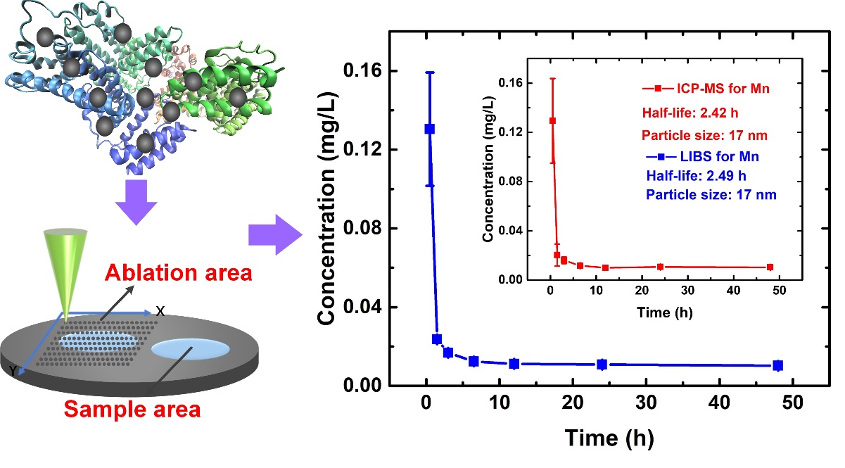
Fig1:Nano-drug half-life detection process
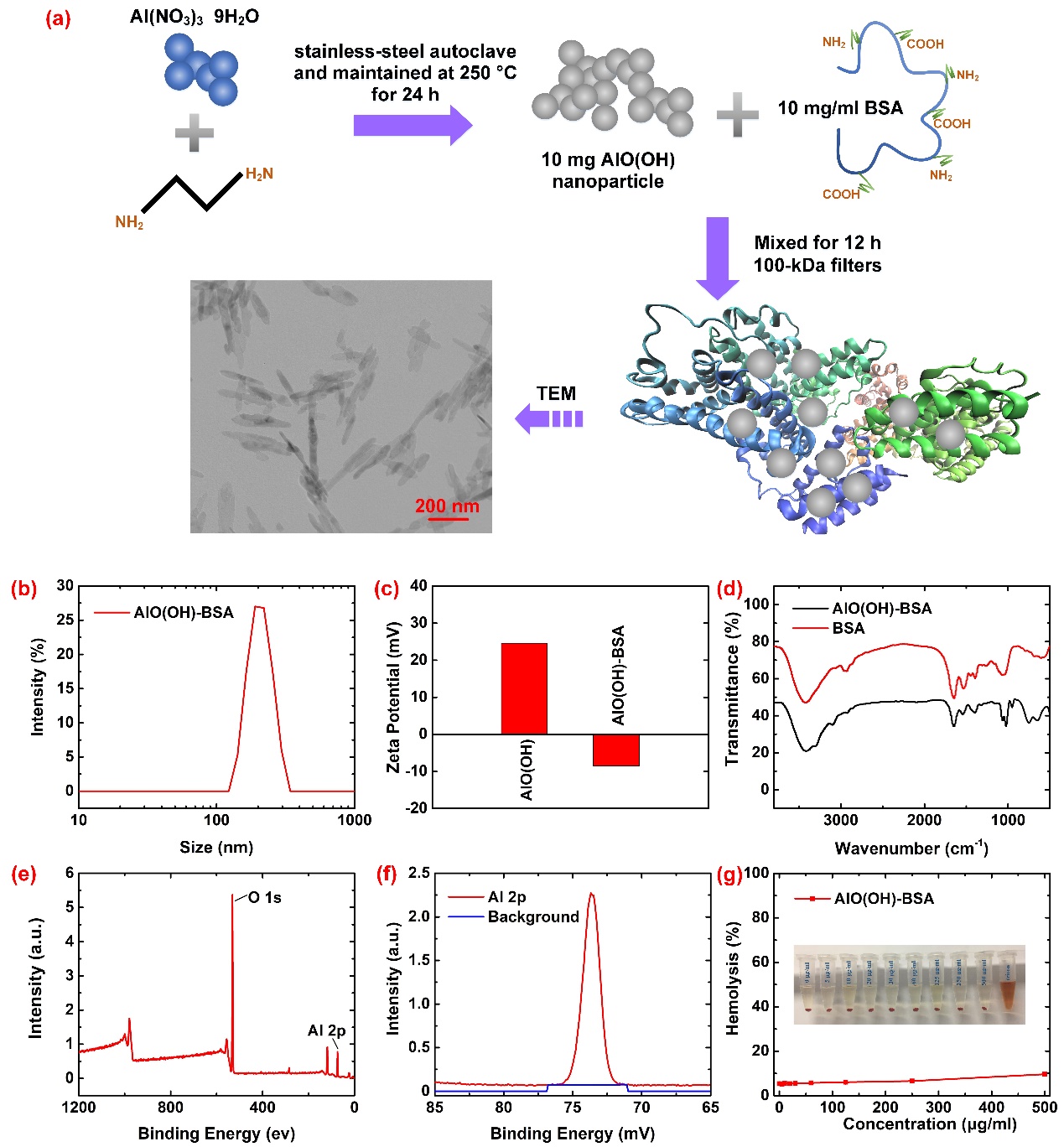
First, researchers synthesized and characterized MnO2-BSA and AlO (OH)-BSA materials. Figure 2 (a) shows the schematic diagram of the synthesis process and included the results of SEM inspection. Figure 2 (b-d) are the characterization results of dynamic light scattering, zeta potential, and near infrared absorption spectrum, respectively. Figure 2 (e-f) are the results of X-ray photoelectron spectroscopy. Figure 2 (g) shows the results of the hemolysis test indicating strong biocompatibility. After MnO2-BSA and AlO (OH) -BSA were successfully synthesized, they were injected into mice for animal experiments, and blood samples were collected at various time points.

Fig2:Synthetic diagram and characterization results for AlO(OH)-BSA
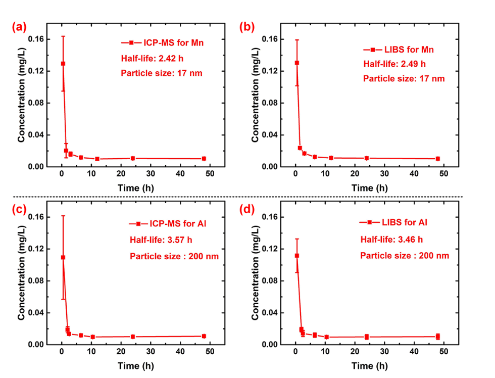
Then, blood samples were prepared for blood collection, including digestion, titration, heating and other operations. After the laser-induced breakdown spectrum acquisition was completed, polynomial fitting and Lorentz fitting were used to remove spectral interference and background. Then the support vector machine regression model based on RBF kernel function was used for quantitative analysis of the corrected spectrum. Finally, the quantitative analysis results were compared with the ICP-MS results, and the relative error was less than 5%.

Fig3: The detection results of LIBS and ICP-MS
The research work was supported by the National Key R & D Program (No: 2018YFA0702100). Associate Professor Guo Lianbo and Professor Jiang Fagang are co-corresponding authors. The laser photoacoustic team of the Wuhan National Optoelectronic Research Center and the Jiang Fagang / Jin Honglin research team completed in collaboration. Thanks to the Cancer Hospital of Union Hospital for its strong support in sample preparation and animal experiments.