Expansion microscopy can overcome the resolutionlimit of traditional optical microscopy by physical expansion of the biological sample embedded with special swellable hydrogels.It enables conventional optical microscopy to observe biological samples athigher resolution (super-resolution). Since first proposed by Boyden et al. in 2015, Expansion microscopy has been extensively developed. At present, most of the polymer hydrogels used for expansion microscopy are obtained by copolymerization of acrylamide, sodium acrylate and covalent crosslinkers (e.g., N, N'-methylenebisacrylamide). Thus the preparation and composition of the hydrogels are relatively complex. If homopolymers can be used to realize tissue embedding and expansion, expansion microscopy research and application will be more convenient and efficient.

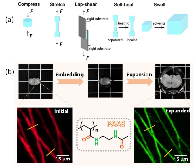
Figure 1.(a) Schematic diagram of the mechanical and swelling properties of PAAE hydrogel; (b) The molecular structure of PAAE, the process of embedding and expansion of mouse brain slices, and the fluorescence images before and after expansion
Recently, Zhu, Li, Luo andcoworkers designed and synthesized a small molecule monomer N-(2-acetamidoethyl)acrylamide (AAE) with a bisamide structure, which polymerized at 40°C to form poly[N-(2-acetylaminoethyl)acrylamide] (PAAE) homopolymer supramolecular hydrogel (Figure 1). There are severalkindof hydrogen bond cross-linking points with different strengths in the hydrogel network structure, which endows the hydrogel with reversible tension/compression, self-healing and adhesion properties. The strong hydrogen bond cross-linking domains in the PAAE hydrogel network structure can remain stable in water and not be dissociated, while the weak hydrogen bond clusters can be dissociated. Therefor,PAAE homopolymer hydrogel can stably swell in water to achieve an equilibrium swelling ratio (calculated by mass) of 1000%. Based on the good biocompatibility and high optical transparency of PAAE hydrogel, PAAE hydrogel was successfully used to realize the embedding and uniform expansion of mouse brain slices with preserved structure integrity (Figure 1). Compared with the fluorescence images before expansion, the fluorescence images of the same brain slice after expansion are composed of more pixels, which is better than the one before expansion. (Figure 1). The two axons that cannot be separated in the image before expansion can be clearly distinguished after expansion, and the resolution is increased to about 1.7 times of the one before expansion. Hence, PAAE hydrogel shows great application prospects in expansion microscopy.
The main innovation of this work is that we provide a new idea, i.e., homopolymer supramolecular hydrogel, for tissue (especially large tissue samples) embedding and expansion microscopy.
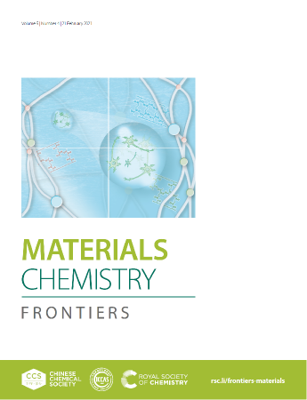
This work was published on Materials Chemistry Frontiers as a hot paper and was selected as the font cover (Figure2). This work was supported by the National Natural Science Foundation of China, the National Basic ResearchProgram (973) of China and the others.
The authors are Peng-Ju Zhao, Chong Li (corresponding author), Ya-Long Wang, Cheng Fan, Xiangning Li, Hui Gong (corresponding author), Qingming Luo and Ming-Qiang Zhu (corresponding author).
Title: Poly[N-(2-acetamidoethyl)acrylamide] supramolecular hydrogels with multiple H-bond crosslinking enable mouse brain embedding and expansion microscopy
Full text link:https://doi.org/10.1039/D0QM00733A

Figure2.Thisworkwas selected as the font cover ofMaterials Chemistry Frontier