On 8th July 2019, in the journal Nature Neuroscience, Dr. Luo Qingming’s team at Huazhong University of Science and Technology published their most recent work “A whole-brain map of long-range inputs to GABAergic interneurons in the mouse medial prefrontal cortex”, which provided the most comprehensive atlas to date of long range inputs to three types of GABAergic neurons in medial prefrontal cortex (mPFC).
GABAergic neurons in the medial prefrontal cortex (mPFC) consist of several sub-groups, including parvalbumin-expressing (PV+), somatostatin-expressing (SST+) and vasoactive intestinal peptide-expressing (VIP+) neurons. They play an important role in regulating working memory, decision-making and emotion associated with motivational and aversive behaviors and their dysfunction has been implicated in various diseases, including major depression, schizophrenia and epilepsy. To better understand the organization pattern and specific functions of mPFC circuits, one essential step is to determine where interneurons receive the information from, especially the long-range direct synaptic inputs at the whole brain level.
Dr. Luo Qingming’s team used monosynaptic rabies viral tracers in combination with fluorescence micro-optical sectioning tomography system (fMOST) to generate a whole brain atlas of the direct long range inputs to GABAergic interneurons in mPFC. They discovered three subtypes of GABAergic interneurons in two areas of mPFC that innervated by same upstream areas, with quantitative differences. In subcortical upstream areas, multiple neural modulators regulated these interneurons, including cholinergic neurons in the basal forebrain and serotoninergic neurons in the raphe nuclei, which could co-release glutamate.
Fine morphology of input neurons, reconstructed from the high-resolution whole brain dataset, showed new wiring diagrams, including connections among the substantia innominata (SI)-anteromedial thalamic nucleus (AM)-mPFC and connections among striatum-AM-mPFC circuit, which shed light on the classic basal ganglia circuitry. Meanwhile, based on the projection logic of individual neuron, the researchers classified the cortical and hippocampal input neurons into several types. The distinct input neurons may transmit different information to carry out different functions.
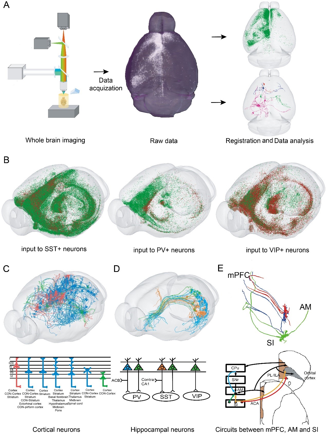
Fig 1 Dissection of long range inputs to three types of GABAergic neurons in mouse medial prefrontal cortex with fluorescence micro-optical sectioning tomography and rabies virus tracing. A. Experimental pipeline. B. The distribution of long range input neurons to three types of GABAergic neurons in mPFC. C. Different cortical input neurons. D. Different hippocampal input neurons. E. New neural circuits among mPFC, SI and AM.
In general, this study generates the long-range inputome of PV+, SST+ and VIP+ neurons in mPFC. This atlas contains information regarding not only the distribution but also the neurochemical properties and fine morphology of input neurons. New types of cortical and subcortical neurons and novel circuits between mPFC and upstream areas were identified. This atlas can facilitate the modeling and understanding of the functional differences of different GABAergic neurons in mPFC, which may, in turn, shed light on the treatment of mental disorders associated with mPFC dysfunction.
This work was mainly completed by PhD student Qingtao Sun and Associate Professor Xiangning Li. Miao Ren, Mengting Zhao, Qiuyuan Zhong, Pan Luo, Hong Ni, Xiaoyu Zhang, Chen Zhang, Jing Yuan, Anan Li and Professor Hui Gong also contributed to this work. Dr. Minmin Luo and Yuqi Ren from National Institute of Biological Sciences helped with the electrophysiology recordings. This work was funded by National Natural Science Foundation of China (fund No. 31871088,91632302,61721092).