The human brain has about 100 billion neurons, through countless synaptic connections they constitute the genius brain network and perform the diverse brain functions such as emotion, memory, and movements.
Classify the neuron types that exist in the brain network and reveal their connection patterns and functional mechanisms is one of the highest priority for the neuroscience study, and it has been taken as the most fundamental part in the diverse big brain projects (ie. United States BRAIN Initiative, The Human Brain Project).
In the cerebral cortex, under the regulation of GABAergic interneurons, pyramidal neurons (PyNs) conducted the main information storage, processing, and transmissions. Among all the interneuron types, AACs (also known as chandelier cells, ChCs) specifically innervate the pyramidal axon initial segments (AISs) where action potential was generated, thus are thought to be playing the most powerful role on controlling PyN activities. And it is highly related with brain diseases such as schizophrenia and epilepsy. Therefore, systematically study the cell types and subtypes of AAC would facilitate our better understanding of its connectivity and function mechanisms in the brain circuits.

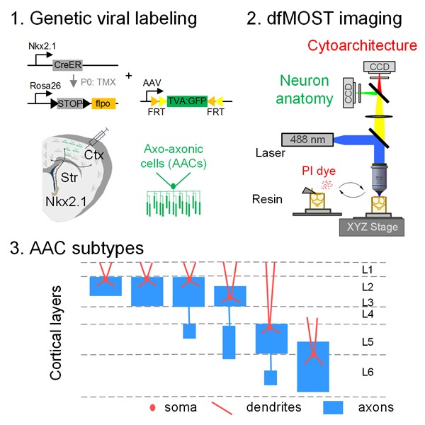
Figure 1. Genetic Single Neuron Anatomy reveals AAC subtypes.
To this goal, researchers of the MOST team lead by Prof. Qingming Luo from the Wuhan National Laboratory for Optoelectronics, Huazhong University of Science and Technology, collaborate with the Huang Lab members lead by Prof. Z. Josh Huang from Cold Spring Harbor Laboratory have designed a genetic Single Neuron Anatomy (gSNA) platform that enables the systematic labeling, imaging, reconstructing, and analyzing of the single AAC morphologies at the fine and complete level.
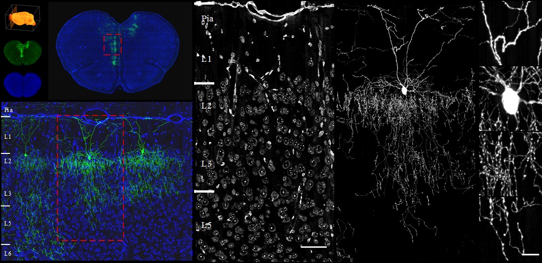
Single neuron anatomy can intuitively indicate the input-output characteristics of a specific neuron. The MOST team used the fMOST system to obtain the high-quality of original image dataset at the whole brain scale, which is especially necessary for the afterward complete reconstruction of the densely-branched single AACs. And the imaging can also capture the whole brain cytoarchitecture information, ensuring the accurate discrimination of brain regions and quantitative analysis (Figure 2).

Figure 2. Whole-brain fMOST imaging result on sparse labeled AACs.
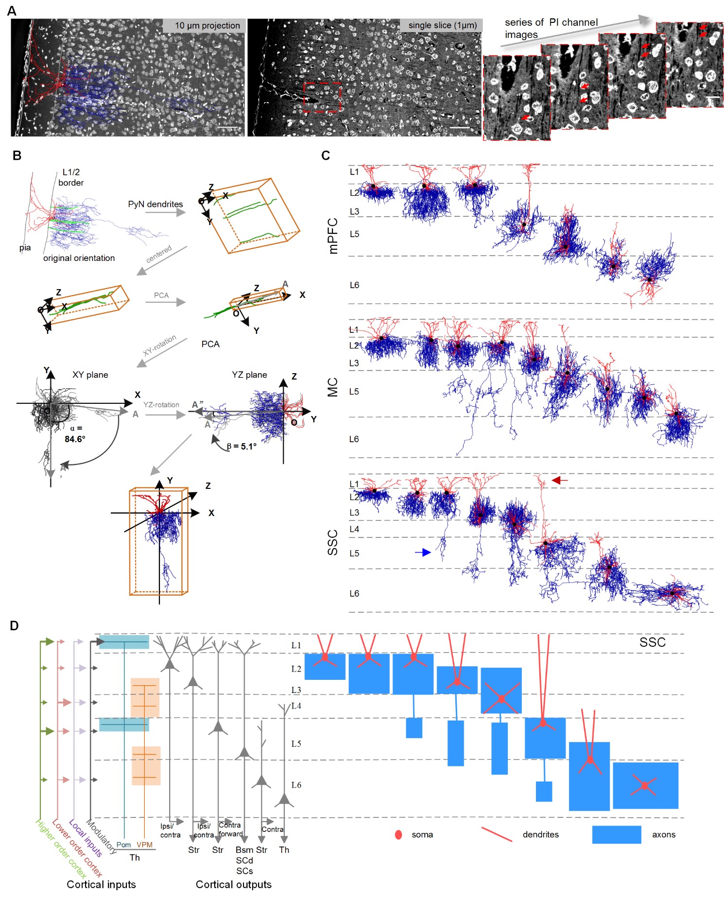
Researchers have found that AACs are not randomly distributed, but are remarkably specific in their location, shape, and purpose. Thus, the collaboration team did a systematic single neuron reconstruction in the neocortical mPFC, MC and SSC areas (Figure 3). Through quantitative analyzing their laminar positions and dendritic and axonal arborization patterns, they determined that there are a wide variety of AAC subtypes which are laminar organized and integrated in the brain network (Figure 1). The new AAC cell subtypes found in the complete anatomical study of single neurons further updated our knowledge on AACs and would be very helpful for the future study on how AACs connected and functioned in the normal and diseased neuronal circuits.

Figure 3. Single AAC reconstructions in the neocortex
This collaboration work has been published in the Cell Reports journal entitled "Genetic Single Neuron Anatomy Reveals Fine Granularity of Cortical Axo-Axonic Cells[1] ".
It is reported that this work was supported by the National Natural Science Foundation of China (61721092 and 81827901), and the Director Fund of Wuhan National Laboratory for Optoelectronics. Prof. Qingming Luo and Prof. Z. Josh Huang are co-corresponding authors of the paper. Dr. Xiaojun Wang is the first author of the paper. Dr. Jason Tucciarone from the Laboratory of Cold Spring Harbor, USA and PhD. student Siqi Jiang from Huazhong University of Science and Technology are the co-second authors. Fangfang Yin, Yao Jia, Xueyan Jia, Yuxin Li, Tao Yang, Zhengchao Xu, Prof. Shaoqun Zeng and Prof. Hui Gong from Huazhong University of Science and Technology participated as co-authors. The article collaborators also include researchers from the Ohio State University, George Mason University.
Reference
[1] Wang, X., Tucciarone, J., Jiang, S., et al. Genetic Single Neuron Anatomy Reveals Fine Granularity of Cortical Axo-Axonic Cells. Cell Reports, 2019, 26 (11): 3145-3159 e5.