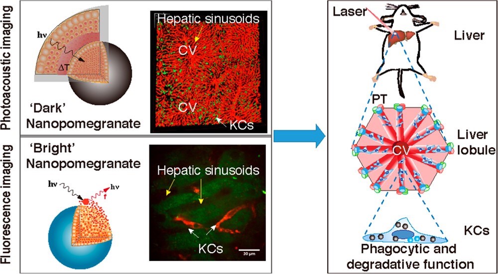
On February 4, 2019, ACS Nano published the latest research results of Professor Zhihong Zhang group from the Wuhan National Laboratory for Optoelectronics of Huazhong University of Science and Technology. They reported a self-assembled “off/on” nano-pomegranate for in vivo photoacoustic/fluorescence bimodal imaging and presented the strategic arrangement of Kupffer cells (KCs) in mouse liver.(Self-Assembled “Off/On” Nanopomegranate for In Vivo Photoacoustic and Fluorescence Imaging: Strategic Arrangement of Kupffer Cells in Mouse Hepatic Lobules. ACS Nano, 2019, 13(2):1526-1537 https://doi.org/10.1021/acsnano.8b07283 )
Macrophages developed from yolk sac are widely distributed in various tissues and organs of the body. They can phagocytose and kill parasites, bacteria, dead cells and tumor cells, participate in the body's immune defense, immune homeostasis and immune regulation. As an important part of the immune system, KCs are macrophages distributed in the liver and play a key role in clearing pathogens and other particulate matters circulating throughout the body. Although people have long known about KCs, its arrangement in the hepatic lobules and its phagocytic degradation ability are still unclear. Liver imaging with antibodies as KCs tracers is often difficult to achieve better imaging depth, field of view and spatial resolution. It seems impossible to simultaneously acquire the basic structural elements of the liver, the fine structure information of the hepatic lobules, and the accurate localization and functional information of the KC.
Photoacoustic imaging combines the advantages of high contrast in optical imaging and high penetration in ultrasound imaging. Photoacoustic imaging has high resolution (lateral resolution 1-2 μm), large field of view (2 mm *2 mm) and relatively deep tissue penetration (300 μm).These advantages make it suitable for deep imaging for the liver. Profs. Zhihong Zhang and Qingming Luo have been studied on the regulation and function of KC of the liver. In their group, doctoral candidate Deqiang Deng and postdoctoral researcher Qiaoya Lin constructed a particle that could be self-assembled to form a pomegranate-like structure. In this work, a kind of hydrophobic near-infrared fluorescent dye could be efficiently self-assembled into tens of thousands of pomegranate seeds (4-5 nm). Then the pomegranate seeds formed a nano-pomegrante with a particle size of 400 nm, whose fluorescence signal was highly quenched (99.74%).When the nano-pomegranate structure remains intact, the fluorescent dye has extremely strong optical absorption characteristics, and it is called "Dark nano-pomegrante"; when the nano-pomegranate-like structure is loose or degraded, the fluorescence quenching effect is weaken, and the fluorescent signal is released, and this particle is called "Bright Nano-pomegranate". Due to its particle size of hundreds of nanometers, it can be rapidly taken up by KCs in the liver after intravenous injection, and the uptake rate is as high as 98.8%. Therefore, the absorption signal of "dark nano-pomegranate" in the liver can be detected by photoacoustic imaging, which can reflect the phagocytic ability of KC. By detecting the fluorescent signal of "light nano-pomegranate" in the liver, the degradation function of KCs can be indicated.

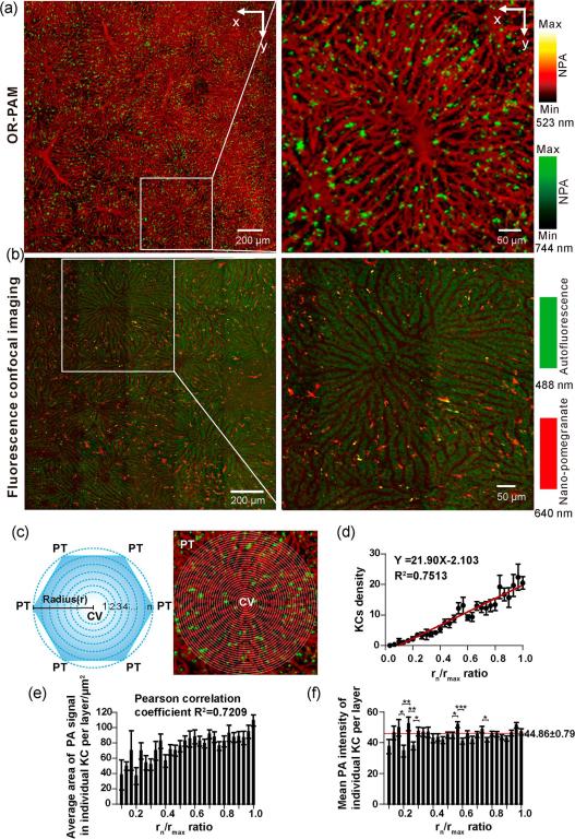
Photoacoustic imaging of the living liver clearly showed the fine structures of the hepatic lobules and hepatic sinusoids in a crisscross pattern with a diameter of 8.4 ± 2.28 μm. Dual-wavelength photoacoustic imaging (523 nm and 744 nm for detecting the absorption signals of hemoglobin and nano-pomegranate, respectively) showed that the distribution density of KCs in hepatic lobules was closely related to its spatial location, which is expressed as the distance from the central vein.. There was a linear relationship between the density of KCs in a single hepatic lobule and its distance from the central vein (rn/rmax) (linear regression coefficient of determination R2 = 0.751). In order to characterize the relationship between the phagocytic function of KCs and its spatial position, we quantitatively analyzed the photoacoustic signal area and intensity of a single KC. The results showed that the photoacoustic signal area of KC is linearly related to its spatial position (rn/rmax) (Pearson correlation the coefficient R2 = 0.7209); and the photoacoustic signal intensity of KC exhibited Zig-zag-like fluctuations only in the region closer to the central vein (rn/rmax: 0.167-0.3) in the hepatic lobules .The intensities of the KC photoacoustic signals at the neutral and distal locations are similar, which may be related to the blood flow velocity in different regions of the hepatic sinusoids. Fluorescence microscopy imaging of living liver showed that KCs released fluorescence slowly (2 h) after phagocytosis of nano-pomegranate, compared with commercial FluoSpheres, and the morphology of KCs was more clearly demonstrated. There are almost no difference of fluorescence intensity between KCs at different spatial positions, and the degradation function of KCs was not affected by its spatial localization.
In summary, the study successfully solved the problem of high-efficiency targeted KCs and simultaneous photoacoustic/fluorescent bimodal imaging, revealing the strategic arrangement and function of KCs in living liver, and expanding our understanding about the physiology of liver. The research work was funded by the National Science Fund for Distinguished Young Scholars (Grant No.81625012), the National Natural Science Foundation of China (Grant No.91442201) and the National Natural Science Foundation of China (Scientific Research Fund Science Foundation (Grant No.61721092). Profs. Zhihong Zhang and Qingming Luo are the co-corresponding authors of this paper. Doctoral candidate Deqiang Deng and postdoctoral researcher Qiaoya Lin are the co-first authors. As co-authors, Prof. Yang Xiaoquan and Doctoral candidates Xianlin Song and Bolei Dai participated in the related work
Full text link: https://pubs.acs.org.ccindex.cn/doi/abs/10.1021/acsnano.8b07283

Fig.Photoacoustic/fluorescent images of KC in mouse hepatic lobules
a) Dual-wavelength photoacoustic imaging of hepatic sinusoids (523 nm, red) and KCs (744 nm, green) in living livers obtained using the OR-PAM system; b) KCs (640 nm, red) 2 hours after intravenous injection of nano-pomegranate Confocal fluorescence imaging with hepatic lobules autofluorescence (488 nm, green); c) Schematic diagram of calculating KCs density in hepatic lobules, 8 μm per layer (rn-rn-1 = 8 μm), and rn / rmax from the central vein The ratio is expressed; d) the correlation between the KC density of each layer and the rn / rmax ratio. e) Quantitative data of the average area of the PA signal for a single KC in each layer and f) the ratio of the average PA intensity to the rn / rmax ratio of a single KC in each layer.