Photoacoustic microscopy (PAM), by combining high contrast based on optical absorption andsensitiveultrasonic detection, has shown great potential in various biomedical applications including tumorangiogenesis, drug effect assessing and brain activities. As an important branch of PAM, optical-resolution PAM can provide high spatial resolution, however suffers fromlimiteddepth of field (DoF), which led to slow volumetric imaging speed and non-uniform imaging quality for biological tissues with large-volume.
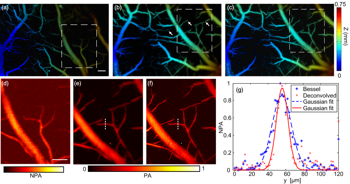
The research team led by Prof. Qingming Luoimplemented a reflection-mode Bessel-beamphotoacoustic microscope with a lateral resolution of 1.6μmand a DoF of 483μm, by using an axicon and an annular mask.The cerebral vasculature of a mouse was imagedin vivoand blind deconvolution was employed to improve the image quality. The results were the first demonstration ofin vivoapplication of a Bessel-beam PAM. This system can be probably used to obtainin vivoimaging of biological tissues subject to breathing movement, thus broaden the application range of PAM.
OnAugust23rd, this work was published in OpticsExpress(Vol.28, No. 18, pp.20167-20176, 2016). The work was supported by Science Fund for Creative Research Group of China (Grant No. 61421064),National Natural Science Foundation of China (NSFC) (Grants No. 91442201)andDirector Fund of WNLO.

The image shows thein vivoimaging results of a mouse cerebral vasculature obtained by a Gaussian beam PAM and a Bessel beam PAM, more vessels can be revealed by the latter one.