Hybrid fluorescence molecular tomography and X-ray computed tomography (FMT-XCT) can noninvasively resolve the three-dimensional (3D) spatial distribution of fluorescent markers and therefore plays an important role inthein vivostudy of physiological and pathological processes of small animals.
When CCD-based free-space fluorescence molecular tomography (FMT) is usedfor imaging of fluorescent targets with a large concentration difference, the limited dynamic range of the CCD diminishes the localization and quantitative accuracy of FMT.
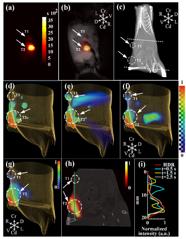
To overcome this,Qingming Luo’s group from Britton Chance Center for Biomedical Photonics, Wuhan National Laboratory for Optoelectronicspresented a high-dynamic-range FMT (HDR-FMT) method. Under the multiple-exposure scheme, HDR fluorescence projection images are constructed using the recovered CCD response curve. Image reconstruction is implemented using iterative reweighted L1 regularization which can reduce artifacts by using fewer HDR fluorescence projection images. Phantom andin vivoanimal studies indicate that localization of fluorescent targets with a large concentration difference is effectively improved with HDR-FMT and with good quantitative accuracy.
The research article “High-dynamic-range fluorescence molecular tomography for imaging of fluorescent targets with large concentration differences” was published online inOptics Expresson 19 Aug 2016 (Vol. 24, Issue 17, pp. 19920-19933). This work is supported by the Key Research and Development Program (2016YFA0201403), Science Fund for Creative Research Group (61421064), National Natural Science Fund (91442201, 61078072), andFundamental Research Funds for the Central Universities (0118187124).
(links to the papers: https://www.osapublishing.org/oe/abstract.cfm?uri=oe-24-17-19920)

Fig.1. Comparison of the results of the tumor-bearing mouse study. (a)Fluorescence reflectanceimage. (b) Fluorescence reflectance image superimposed on a white light image.Tumors are labeled T1 and T2. (c) 3D rendering of the mouse and highlighted tumor areas (in the white dashed circles). Region between the two dashed lines was used for FMT reconstruction. (d)–(f) 3D rendering of mouse skin based on XCT and fluorescence signals based on FMT reconstruction using low-dynamic-range fluorescence projection images obtained with exposure times of0.5,1.5, and2.5s, respectively. (g) 3D rendering of mouse skin and fluorescence signals based on HDR-FMT. (h) Overlay of oblique sections obtained from XCT and HDR-FMT. White arrows point to the reconstructed FMT signals.(i)Intensity profiles of the HDR-FMT and low-dynamic-range reconstructed results. The red, cyan, yellow, and gray lines represent the reconstructed results of HDR-FMT, exposure time of 0.5 s, 1.5 s, and 2.5 s, respectively. [Coordinate system was defined by D (dorsal), V (ventral), Cr (cranial), Cd (caudal), L (left), and R (right). 3D renderings in (c)–(g) were implemented using AMIRA software.]