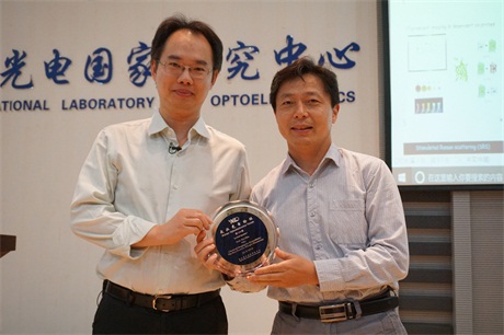
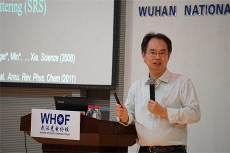
WUHAN, China (May 28, 2018) - Wuhan Optoelectronics Forum No. 139 was successfully held in Auditorium A101 at Wuhan National Laboratory for Optoelectronics (WNLO) in the morning of May 28. Prof. Wei Min from Columbia University delivered an exciting talk entitled Chemical Imaging for Biomedicine: The Next Frontier of Light Microscopy. Prof. Ping Wang chaired the forum. Prof. ShaoQun Zeng, the Academic Advisory Committee Member of WNLO, awarded Prof. Wei Min the forum medal.
Innovations in light microscopy have tremendously revolutionized the way researchers study biological systems. Although fluorescence microscopy is currently the method of choice for cellular imaging, it faces fundamental limitations such as the bulky fluorescent tags and limited multiplexing ability in the era of “omics”. In this talk, I will present two chemical imaging strategies, respectively. First, we devised a live-cell Bio-orthogonal Chemical Imaging platform suited for probing the dynamics of small bio-molecules, which can not be effectively labeled by bulky fluorophores. This scheme couples the emerging stimulated Raman scattering microscopy with tiny and Raman-active vibrational probes (e.g., alkynes, nitriles and stable isotopes including 2H and 13C). Exciting biomedical applications such as imaging fatty acid metabolism related to lipotoxicity, glucose uptake and metabolism, drug trafficking, protein synthesis in brain, DNA replication, protein degradation, RNA synthesis and tumor metabolism will be presented. Second, we invented a super-multiplex optical imaging technique. We developed electronic pre-resonance stimulated Raman scattering (epr-SRS) microscopy, achieving exquisite vibrational selectivity with high versatility and sensitivity. Chemically, we created a unique vibrational palette consisting of novel dyes bearing conjugated and isotopically-edited triple bonds, each displaying a single epr-SRS peak in the cell-silent spectra window. Up to 24 resolvable colors are currently achieved with great potential for further expansion. Using this approach, we monitored DNA and protein metabolism in neuronal co-cultures and brain tissues. This super-multiplex optical imaging approach promises to facilitate untangling the intricate interactions in complex biological systems, and find broad applications in photonics and biotechnology in general.
Dr. Wei Min graduated from Peking University with a Bachelor's degree of Chemistry in 2003. He received his Ph.D. from Harvard University in 2008 studying single-molecule biophysics with Prof. Sunney Xie. After continuing his postdoctoral work in Xie group, Dr. Min joined the faculty of Department of Chemistry at Columbia University in 2010, and is promoted to Full Professor there in 2017. He is also affiliated with the Kavli Institute for Brain Science at Columbia University. Dr. Min's contribution has been recognized by a number of honors, including: 2017 American Chemical Societies(ACS) Early Career Award of Experimental Physical Chemistry、2017 Coblentz Award of Molecular Spectroscopy、2015 Buck-Whitney Award of ACS Eastern New York Section、2015 Camille Dreyfus Teacher-Scholar Award、2013 Alfred P. Sloan Research Fellowship. Dr. Min's current research interests focus on developing novel optical spectroscopy and microscopy technology to address biomedical problems. In particular, his group has made important contributions to the development of stimulated Raman scattering (SRS) microscopy and its broad application in biomedical imaging including bioorthogonal chemical imaging of small molecules and super-multiplex vibrational imaging. Professor Wei has published many scientific papers on Science, Nature, Nature methods and other high impact journals.