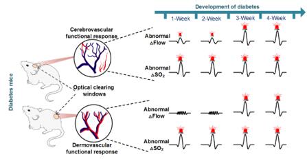
The vascular dysfunction induced by glucose metabolic disorders can induce various complications, and the rates of disability and death for diabetic patients who suffer from stroke are much higher than non-diabetic patients. However, there are no efficient approaches to early warn the cerebrovascular diseases caused by diabetes because diabetes is a chronic and complicated disease. It is hard to obtain cerebrovascular function with high resolution to long-term monitor its changes in clinical or on in vivo animals. Recently, the magazine named “Theranostics” (2018 IF:8.063) in Biomedical scope has reported an article of Prof. Dan Zhu of Wuhan National Laboratory for Optoelectronics in HUST. They firstly realize chronically in vivo monitoring the cerebral and cutaneous vascular dysfunction in diabetic mouse by combining their special skin/skull optical clearing techniques with optical imaging methods, by which they find that the blood oxygen metabolism dysfunction of cutaneous vessels can be an early-warning indicator for predicting the diabetes-induced cerebral vascular dysfunction.

Diabetes can induce various complications, which are mainly caused by the vascular dysfunctions. The skin, as an accessible and generalized vascular bed, has shown great potentials in predicting cardiovascular disease and diabetic retinopathy. Whether the skin can also be a suitable and replaceable vascular bed to predict the diabetes-induced cerebrovascular dysfunction? It needs further investigations to study if there are correlations between cerebral and cutaneous vascular dysfunctions along the development of diabetes.
The development of optical imaging techniques make it possible to monitor the responses of cutaneous/cortical vascular functions in pathophysiological conditions. However, the high scattering of skin and skull strongly hinder the light penetration in biological tissues. Though the skin/skull windows established by surgical operations help to acquire cutaneous/cortical vascular structures and functions with high resolution, but they inevitably cause inflammations and even injures.
In this study, the authors dynamically monitored the cutaneous/cortical blood flow and blood oxygen changes stimulated by sodium nitroprusside and acetylcholine at the different stages of diabetes by combining recently-developed skull and skin optical clearing techniques with laser speckle contrast imaging and hyperspectral imaging system, by which they quantitatively analyzed the cutaneous cortical functional responses caused by medicines, compared their differences then further explored the reason. The results showed that the cutaneous vascular blood oxygen dysfunction happens earlier than blood flow dysfunction, and both the blood oxygen and blood flow dysfunctions of cortical vessels occur at the early stage of diabetes. Therefore, diabetes-induced cutaneous vascular blood oxygen changes has the potential to serve as a good assessment indicator for revealing cerebrovascular dysfunction in the early stage of diabetes. This research has great clinical value for the diagnosis and medical intervention in the early stages of diabetes.

This work was supported by National Natural Science Foundation of China (NSFC) (Grant No.61860206009, 81870934, 31571002, 61721092), the National Key Research and Development Program of China (2017YFA0700501), the Fundamental Research Funds for the Central Universities, HUST (Grant No. 2019KFYXMBZ069; 2019KFYXJJS052), and the Innovation Fund of WNLO. The PhD student Wei Feng is the first author, and his supervisor Prof. Dan Zhu is the corresponding author.
The original link: http://www.thno.org/v09p5854.htm
Feng Wei, Liu Shaojun, Zhang Chao, Xia Qing, Yu Tingting, Zhu Dan. Comparison of cerebral and cutaneous microvascular dysfunction with the development of type 1 diabetes. Theranostics 2019; 9(20): 5854-5868.