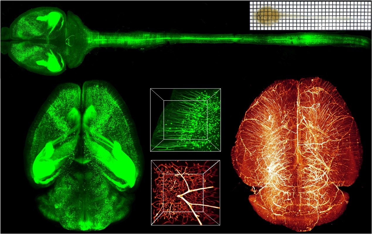
On January 11, 2019, Science Advances published the latest research achievement in Prof. Dan Zhu’s group from Wuhan National Laboratory for Optoelectronics, Huazhong University of Science and Technology titled ‘FDISCO: Advanced solvent-based clearing method for imaging whole organs’.


Figure 1. Three-dimensional imaging and visualization of the neuronal and vascular networks of whole brain by FDISCO.
Tissue optical clearing techniques can render the tissue transparent by reducing the scattering, and improve the light penetration in multiple optical imaging techniques. In recent years, various tissue optical clearing methods have been developed, allowing three-dimensional imaging and reconstruction of the structures of biological tissues with no need of mechanical sectioning, combining with optical microscopies. However, when applied to the large-scale tissues or intact organs, the imaging is still unsatisfactory due to the difficulty to obtain both high-performance clearing and fluorescence-preserving capability.
3DISCO is an organic solvent-based clearing protocol, which can achieve the highest level of tissue transparency in a short time, but can result in a rapid decline in endogenous fluorescence signals during the clearing and storage procedure. To address this issue, Prof. Dan Zhu’s group developed an advanced optical clearing method based on 3DISCO by temperature and pH condition adjustments, named FDISCO. FDISCO can achieve a high level of fluorescence preservation of various probes, such as fluorescent proteins (FPs) and chemical fluorescent tracers, with a short processing time while maintaining potent tissue-clearing capability and tissue shrinkage, thereby facilitating the imaging of large-scale samples. FDISCO allows 3D imaging of neuronal and vascular structures in various samples, including the intact brain, kidney and muscle, in combination with LSFM. Using FDISCO, they detected weakly labeled neurons with virus in the whole brain and analyzed the spatial distribution of cells projecting to the virus-injected regions. FDISCO provides a novel and efficient method for morphological analysis and quantification of anatomical structures of various organs in biological and medical researches.

Figure 2. Repetitive imaging of cleared mouse brain. (a) Fluorescence images of cortical neurons in the FDISCO-cleared brain taken at 0, 150 and 365 days, respectively. (b) The fluorescence level quantification of cleared brains over time after clearing with different methods.
This study was supported by the National Key Research and Development Program of China (grant no. 2017YFA0700501), the National Nature Science Foundation of China (grant nos. 61860206009, 81870934, 31571002, 81701354, and 91749209), the Science Fund for Creative Research Group of China (grant no. 61721092), the Project funded by the China Postdoctoral Science Foundation (grant nos. 2017M612463 and 2018T110772), the Fundamental Research Funds for the Central Universities, HUST (grant no. 2018KFYXKJC026), and the Director Fund of WNLO. Prof Dan Zhu is the corresponding author, Yisong Qi and Tingting Yu are the co-first authors. Jianyi Xu, Peng Wan, Yilin Ma, Jingtan Zhu and Yusha Li are coauthors. Prof. Qingming Luo and Prof. Hui Gong participated in guidance of this work and commented on the manuscript.
Full text link:
http://advances.sciencemag.org/content/5/1/eaau8355