The work of professor Ping Wang from Huazhong University of Science and Technology, China, published online their breakthrough results in Optica(IF:9.778, flagship journal of OSA). This research work demonstrated a novel optical tomography,pulse-sheetchemical tomography (PCT), which allows label-free bond-selective three-dimensional (3D) imaging of large intact tissues. It realizes vibrational tomography of highly scattering bone tissue withlateral resolution of 16.4µm and axial resolution of24.5µm, over a large field of view of 8×8×1.6 mm3 in mouse skull with scalp, providing unique biomedical and clinical prospects for optical tomography of tissues and organs in the future.
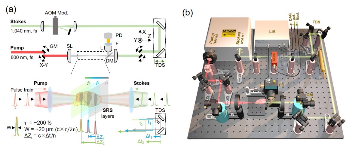
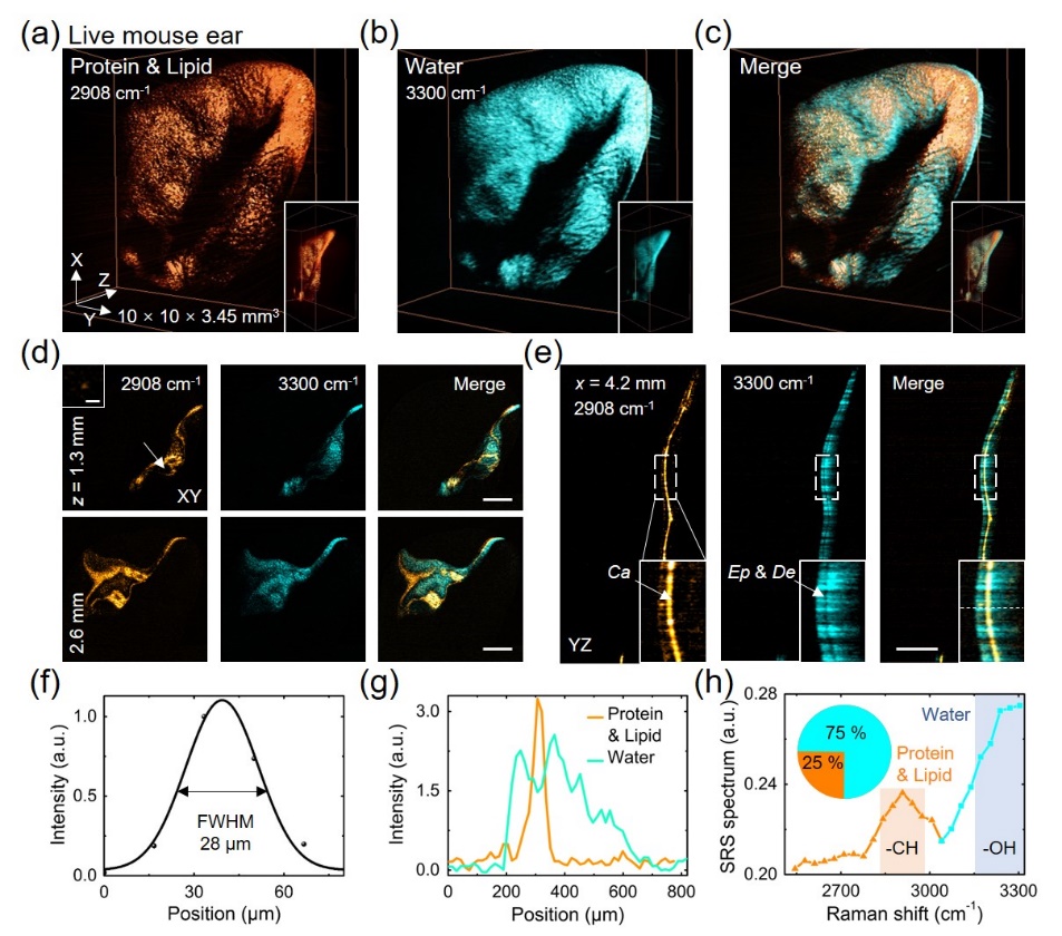
The noninvasive optical tomography with chemical specificity proposed by the team is based on the counterpropagating stimulated Raman scattering effect, which achieves a centimeter-scale and high resolution tomography. By introducing the phase-locked femtosecond pump and Stokes laser pulses into the tissue in a counter-propagating mode,a pulse-duration-determined light sheet forms in the fixed z plane of the tissue as the femtosecond pulse trains repeatedly encounter each other there. Essentially, the three-dimensional (3D) chemical anatomy of the tissue can be achieved by scanning the pulse-sheet plane across the sample by tuning the relative time delay between the pump and Stokes pulses. The PCT substantially relieves the trade-off between optical focal depth and spatial resolution. Although with a low numerical aperture of lens ~0.013, reaching a high axial resolution (~25 μm) in counter-propagating SRS. The team performed large volume imaging of protein components in intact mouse skull and scalp with the system, characterized the difference in thickness distribution of sagittal andparietalbone on the skull, and realized the tomographic imaging of skull and scalp (~400 μm). In addition, the whole ear (10×10 mm2) of living mouse was imaged with PCT system, which revealed the difference of structure and chemical composition between skin and cartilage, and characterized the distribution and proportion of water and protein in the ear. The non-invasive chemical tomography optical imaging technology is very suitable for the chemical composition analysis of large tissue samples. At the same time, the future work is also prospected in the end of this paper. The pulse sheet imaging technology is expected to develop into a new clinical diagnostic technology.

Fig. 1. Pulse sheet chemical tomography imaging system (PCT).a, Principle of pulse sheet system;b, Real experimental schematic diagram of the PCT setup.

Fig. 2. Three-dimensional tomography imaging of intact mouse skull and scalp.

Fig. 3. Three-dimensional tomography imaging of live mouse ear.

Fig. 4.Ping's Lab in Huazhong University of Science and technology.
The Doc. Chi Yang, Yali Bi are the first authors with same contribution. Prof. Ping Wang is the corresponding author. This work is in cooperation withProfessor Zhihong Zhang from Huazhong University of Science and Technology, China.The Wuhan National Laboratory for Optoelectronics of Huazhong University of science and technology is the first unit.
The research work has been strongly supported by National Key Research and Development Program of China (2016YFA0201403); the the National Natural Science Foundation of China (61675075, 61704061, 61974050); Science Fund for Creative Research Groups of China (61421064); Innovation Fund of the Wuhan National Laboratory for Optoelectronics; University of Nebraska-Lincoln Startup Fund.
Article links
C. Yang, Y. Bi, E. Cai, Y. Chen, S. Huang, Z. Zhang, and P. Wang, "Pulse-sheet chemical tomography by counterpropagating stimulated Raman scattering," Optica8, 396-401 (2021).
https://www.osapublishing.org/optica/fulltext.cfm?uri=optica-8-3-396&id=449181###